Skin aging and extracellular vesicles: getting anti-aging actives past the barrier
What aging does to skin, why most actives never reach the dermis, and how vesicles from cells and microbes could carry them there.

October 2026 · 10 min read
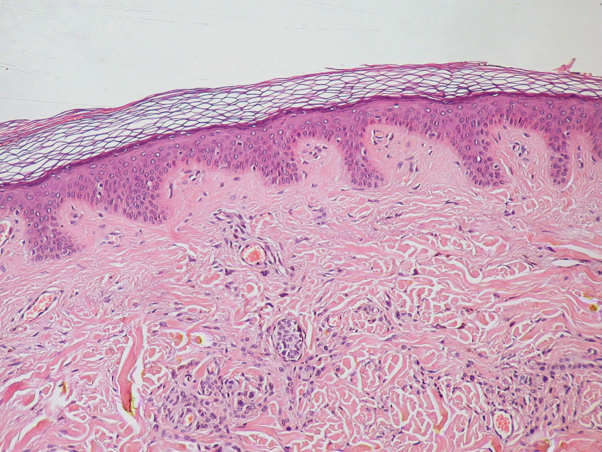
Skin is where aging shows first, and it gets more attention than any other organ when people try to slow aging down. It is also one of the hardest organs to reach. Its outer layer exists to keep things out, so most of what goes into a cream never gets past the surface. This note covers what changes in skin as it ages, why so many active ingredients fail to reach the cells that need them, and why extracellular vesicles, the small particles that cells release to talk to each other, are now the focus of my work.
Three layers, three jobs
The epidermis is the thin outer layer. Its main cells, keratinocytes, divide at the base, move up and flatten as they go, and die at the top. What is left forms the stratum corneum: about 15 to 20 layers of dead, flattened cells held together by lipids, often compared to bricks and mortar. It keeps water in and keeps microbes, irritants and most chemicals out.
Below it sits the dermis, which gives skin its strength and its spring. Fibroblasts in the dermis make collagen, elastin and the gel of sugars and proteins around them. Collagen fibres give skin its resistance to stretching, and elastic fibres let it snap back. Blood vessels, nerves, hair follicles and glands run through the dermis too. Under the dermis is a layer of fat.
Aging changes all three layers, but the dermis matters most for how skin looks and holds up. Wrinkles, sagging and thin, fragile skin mostly come from what happens to its collagen.
Two clocks: time and sunlight
Dermatologists describe two kinds of skin aging. Intrinsic aging is what happens with time alone, and you can see it on skin that has rarely seen the sun, like the inside of the upper arm. Photoaging is the extra damage from ultraviolet light, and it shows on the face, neck and hands.
Fewer fibroblasts, less collagen
In intrinsic aging, the dermis loses cells. A biopsy study of sun-protected skin found about 35% fewer fibroblasts in older adults than in young ones. The fibroblasts that remain make less collagen, and the collagen already there breaks into fragments over time. Reviews of the field describe the result as a net collagen deficit, which shows up as thin skin that tears and bruises easily.
Collagen also lasts a long time. Collagen fibrils in skin have a half-life of about 17 years, so a fibre damaged at 30 can still be there, broken, at 50. Damage builds up much faster than the body clears it.
What sunlight adds
Ultraviolet light speeds all of this up through a family of enzymes called matrix metalloproteinases, or MMPs. Work from Gary Fisher and John Voorhees at the University of Michigan showed that UV exposure switches on MMP-1, MMP-3 and MMP-9 in human skin. MMP-1 makes the first cut in intact collagen, and the other two break the fragments down further. Most of these enzymes come from keratinocytes in the epidermis and diffuse down into the dermis.
UV also stops new collagen from being made. In their experiments, a single moderate dose of UV, enough to turn skin slightly red, nearly shut down the production of new collagen for about a day. Each such exposure breaks down collagen and pauses its replacement. UVA, which reaches deeper into the skin than UVB, is thought to drive much of the long-term damage.
The same group showed that treating skin with retinoic acid before UV exposure blocks much of the rise in MMPs. That is one reason prescription retinoids remain the benchmark that any new anti-aging ingredient has to be measured against.
Senescent cells
Skin also collects senescent cells with age, in the epidermis and in the dermis. A senescent cell has stopped dividing for good but stays alive and keeps secreting. What it secretes, called the senescence-associated secretory phenotype, includes inflammatory signals and the same MMPs that cut collagen. Even a modest number of senescent fibroblasts can therefore add to collagen loss in the tissue around them.
Senescence is not purely harmful. Senescent cells appear briefly during wound healing and help it along. The trouble starts when they stay. My postdoc work on senescence in breast cancer cells, and an earlier note on cells that refuse to die, cover the biology in more detail.
The barrier problem
Repairing the dermis means getting something into it, and the outer layer is built to stop that.
The stratum corneum is very good at its job. Small molecules that dissolve in fats can slowly diffuse through the lipid mortar. Large molecules, charged molecules and most proteins mostly stay on top. That excludes much of what you would want to deliver to an aging dermis: growth factors, peptides, RNA and many plant compounds.
Formulators have tried to get around this for decades with lipid vesicles. Conventional liposomes turned out to be too rigid to cross the stratum corneum. They tend to sit in its upper layers and release their contents slowly from there. Flexible vesicles made with ethanol or surfactants get further, and how far is still debated.
Extracellular vesicles face the same barrier. Some studies of vesicles applied to the skin find them mostly in the stratum corneum, where they may still act on the cells just below. Others report labelled vesicles reaching deeper layers of lab-grown or mouse skin, though often in images rather than measured amounts. Whether intact vesicles cross healthy human skin, and in what numbers, is still an open question. Any serious product in this area has to answer it with data.
What extracellular vesicles are
Every cell releases extracellular vesicles. They are wrapped in a lipid membrane like the cell's own, far too small to see in an ordinary light microscope, and they carry proteins, lipids and RNA from the cell that made them. Cells use them to send signals to neighbours and to distant tissues. Exosomes are one type, formed inside the cell before release, but most preparations hold a mix of types, and "extracellular vesicles" is usually the more accurate term.
Humans, plants, bacteria and yeast all make them. That matters for skin for two reasons. First, vesicles carry signals of their own, so a vesicle preparation can act on skin cells even before anything is loaded into it. Second, their membrane protects what is inside, so they can carry fragile molecules that would break down in a cream or on the skin surface.
The field has learned to be careful with these particles. The International Society for Extracellular Vesicles publishes guidelines, now MISEV2023, on how to isolate vesicles and what to measure before calling something a vesicle preparation: particle size and number, markers that should be present, contaminants that should be absent. Those guidelines exist because early studies often could not tell vesicles from protein clumps and debris that came along during isolation.
Two ways vesicles could help aging skin
Vesicles as the active
The first approach treats the vesicles themselves as the ingredient. Much of the early evidence comes from vesicles made by probiotic bacteria.
In a 2023 study, vesicles from Lactobacillus paracasei were added to human dermal fibroblasts and keratinocytes inflamed with TNF-α, a signal that rises in aged and damaged skin. At low doses, the vesicles restored fibroblast growth and collagen production and lowered MMP-1 and the inflammatory signals IL-6 and IL-8. The authors confirmed the effect in a three-dimensional model of human skin grown in the lab. There was no test in people.
A 2022 study of vesicles from Lactobacillus plantarum did include people. Sixteen women completed a four-week test, applying either the vesicles or a placebo twice a day. The vesicle group showed less wrinkle depth around the eyes and better elasticity, moisture and skin density by instrument measurements. It is a small, short study, and the paper does not say whether it was randomized or blinded. It is a reason to run a better trial, and not yet proof that the vesicles work.
Vesicles as the carrier
The second approach uses vesicles to carry something else: an active ingredient that works on skin cells but cannot reach them unaided. The vesicle protects the cargo from breaking down, and its membrane can fuse with or be taken up by skin cells in ways that a free molecule cannot.
This is the approach behind ProbioVesicle, which I develop at Nanomère, the company I co-founded in May 2026. ProbioVesicle uses vesicles from a probiotic yeast to carry anti-aging actives into the skin.
Yeast is a practical choice. It is food safe, it has a long history in fermentation, and it grows under controlled conditions at scale, which makes it easier to produce vesicles batch after batch with the same properties. Consistency is one of the hardest problems in this field. Yeast is also the organism in which I spent my PhD studying aging. Our screen in Vladimir Titorenko's lab at Concordia found plant extracts that extend the lifespan of yeast, and the most potent, a white willow bark extract called PE21, works by changing how cells handle their lipids. Compounds like that act on aging biology in a dish, but they only matter in people if they reach the right tissue, and in skin that means getting past the barrier.
How the work gets tested
The same logic runs through the vesicle carriers in Christopher Brett's lab at Concordia, where the first carrier holds a chemotherapy drug, and through the skin work. A carrier has to pass a series of tests, and each one can end the project.
First, loading: how much of the active ends up inside the vesicles, measured directly, and whether it stays there. Second, uptake: whether target cells take up the loaded vesicles and whether the cargo gets inside them. Third, effect: whether the loaded vesicle does more than the same active applied on its own, in the same assay. A vesicle that performs no better than the free active adds cost without adding anything.
For skin, two more questions matter. One is depth: where the vesicles end up in a skin model, and in what amounts. The other is the immune response. Skin is full of immune cells, and a carrier that triggers inflammation would make aging worse, since inflammation is one of the things that drives it. Testing how human immune cells respond to a carrier is part of the work from the start; in the Brett lab, that testing is done with partner labs in glycoengineering and human immunology.
Where the evidence stands
Vesicles in skin care are still early science that moved onto store shelves quickly. Products labelled "exosome" now range from careful preparations with published characterization to vials whose contents nobody has measured. An earlier note on exosomes in skincare covers how to read those labels.
Regulators are watching the medical end of this. The US Food and Drug Administration says no exosome product is approved to treat any disease, and it issued a safety alert in 2019 after patients in Nebraska had serious adverse events from unapproved exosome products. Cosmetics are allowed to claim effects on appearance, not on disease, and that line will shape what skin products can say.
The field now needs vesicle preparations characterized to the MISEV standard, studies that measure where the vesicles go in human skin, head-to-head comparisons with the free active, and controlled human studies that are randomized, blinded and long enough to see a change in collagen. The products that last will be the ones that can show those data.
Why this is the focus
Aging research has found many compounds that slow aging in yeast, worms or mice. Far fewer reach a tissue in people at a dose that does anything. Skin is the most accessible place to close that gap: its aging is visible and measurable, its biology is well mapped, and topical products can be tested on people without the risks of a drug that enters the bloodstream. If vesicles can reliably carry actives past the barrier, skin is where that will be shown first.
More on the research behind this is on the research page, and in the notes on aging and drug delivery.