A cell state that is 3% of a precancerous lesion decides whether lung tumours progress
Tracing and killing a high-plasticity cell state in mouse lung adenocarcinoma stalled tumours and reversed resistance to chemotherapy and a KRAS inhibitor.

October 2026 · 3 min read
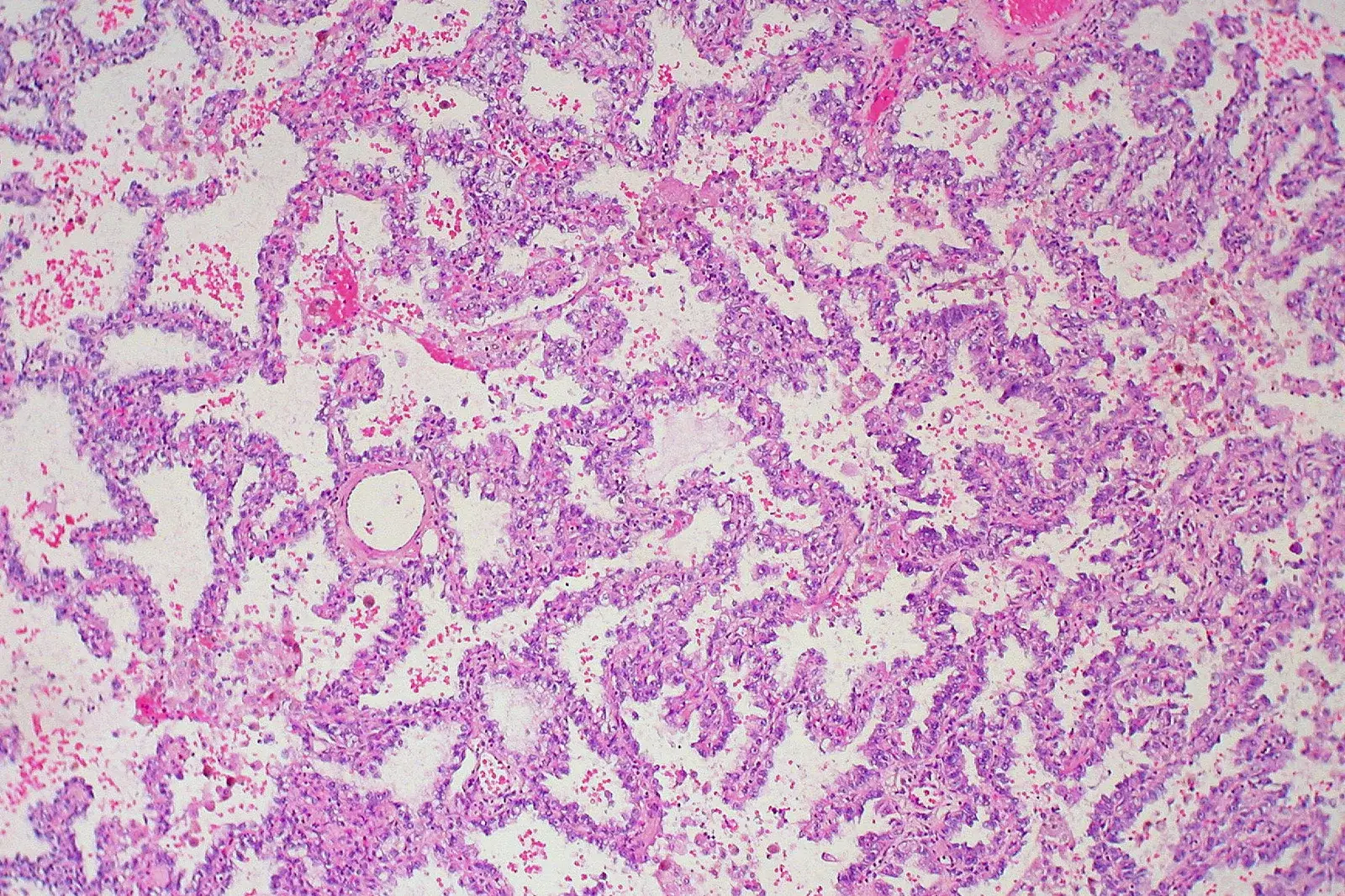
About 3% of cells in a precancerous lung lesion sit in a state that lets them become almost anything else. By the time the tumour is established that fraction is around 15%, and in metastases it reaches 30%. Delete those cells in mice and tumours stall, shrink, and lose their ability to shrug off drugs.
That is the finding reported on 21 January 2026 in Nature by Tuomas Tammela's lab at the Sloan Kettering Institute at Memorial Sloan Kettering, with Jason Chan and Chun-Hao Pan as co-first authors. The paper is a functional test of a cell state the same group described years earlier, and functional tests are where most cell-state stories fall apart.
Labelling a state, then removing it
The team built mouse lung adenocarcinoma models in which the high-plasticity cell state, HPCS, could be detected, lineage-traced and killed on command. Two independent ways of killing it were used: a suicide gene, and CAR T cells directed at uPAR, a surface protein the state carries but most healthy cells do not.
Three results follow from that design:
- HPCS cells are dedifferentiated, and they give rise to both early and advanced cancer cell states. Lineage tracing shows the traffic running outward from them.
- Ablating HPCS slowed progression and reduced tumour burden. Ablating a differentiated cancer cell state did nothing.
- Killing HPCS cells very early prevented mutated cells from progressing to cancer at all, while removing them from established tumours caused significant shrinkage.
The last pair is the one I keep returning to. The same intervention works at two very different points in tumour history, which argues the state is load-bearing throughout rather than a late adaptation.
The decision that creates drug tolerance
Under chemotherapy or a KRAS inhibitor, HPCS cells were the source of the cells that survived, and ablating the state reversed that resistance. This reframes drug tolerance as a question about which state a cell occupies when the drug arrives, rather than which mutation it happens to carry.
It is also a different claim from the classical cancer stem cell. HPCS is not a fixed rare subpopulation. It is a state cells enter in response to injury-like signals, borrowing a regenerative programme that normal epithelium uses to repair itself. The same state appears in regenerating epithelia and in carcinomas of other tissues, which matters because carcinomas account for the large majority of human cancers.
If plasticity is a state rather than a lineage, then the therapeutic target is the entry route into it, not a cell to be counted.
Where to be careful
Everything here is mouse. Autochthonous models are the best available version of a lung tumour arising in its own tissue, but the paper reports no patient data, and the fraction of HPCS cells in human tumours is not established by this work.
uPAR is a rational target precisely because it is largely absent from healthy cells, and uPAR-directed CAR T cells have been used before to clear senescent cells. That makes a safety question concrete rather than hypothetical: uPAR also appears on cells doing legitimate wound repair, and a therapy that removes plasticity from injured tissue may impair healing. The antitumour response in this paper is described qualitatively, without dose-response data.
Two further limits. The resistance result comes from mouse models treated with one chemotherapy and one KRAS inhibitor, and how far it holds across drug classes is still open. And several senior authors have equity and advisory relationships with companies working on this cell state and on uPAR binders, which is normal at this stage but worth stating.
What I would want to see next
The experiment I want is a human one, and it does not require a new drug. Take paired biopsies from lung adenocarcinoma patients before treatment and at the point of minimal residual disease, then ask with single-cell profiling whether HPCS-like cells are enriched in the residual population and whether their abundance before treatment predicts relapse. If the fraction rises under therapy in patients the way it does in mice, this becomes a biomarker before it becomes a target.
Second, I would like to see whether blocking entry into the state, rather than killing cells already in it, is enough. Ablation is a blunt instrument and the surface marker is shared with healthy repair. If the injury-like signal that opens the door can be identified and closed, that is the safer drug.